µkiss-and-tell: A new method for precision delivery of nanoparticles and small molecules to individual cells
The delivery of experimental materials to individual cells with exactness and exclusivity has long been an elusive and much sought-after ability in biology. With it comes the promise of deciphering many longstanding secrets of the cell. A research team at the Max-Planck-Zentrum für Physik und Medizin, Erlangen led by Professor Vahid Sandoghdar has now successfully shown how small molecules and single nanoparticles can be applied directly onto the surface of cells. In the study, which was recently published in the journal Nature Methods, the scientists describe their technique as a “µkiss” (microkiss) – an easy and cost-effective new method, unlocking new possibilities in single-cell science with a view towards next generation therapeutic applications.
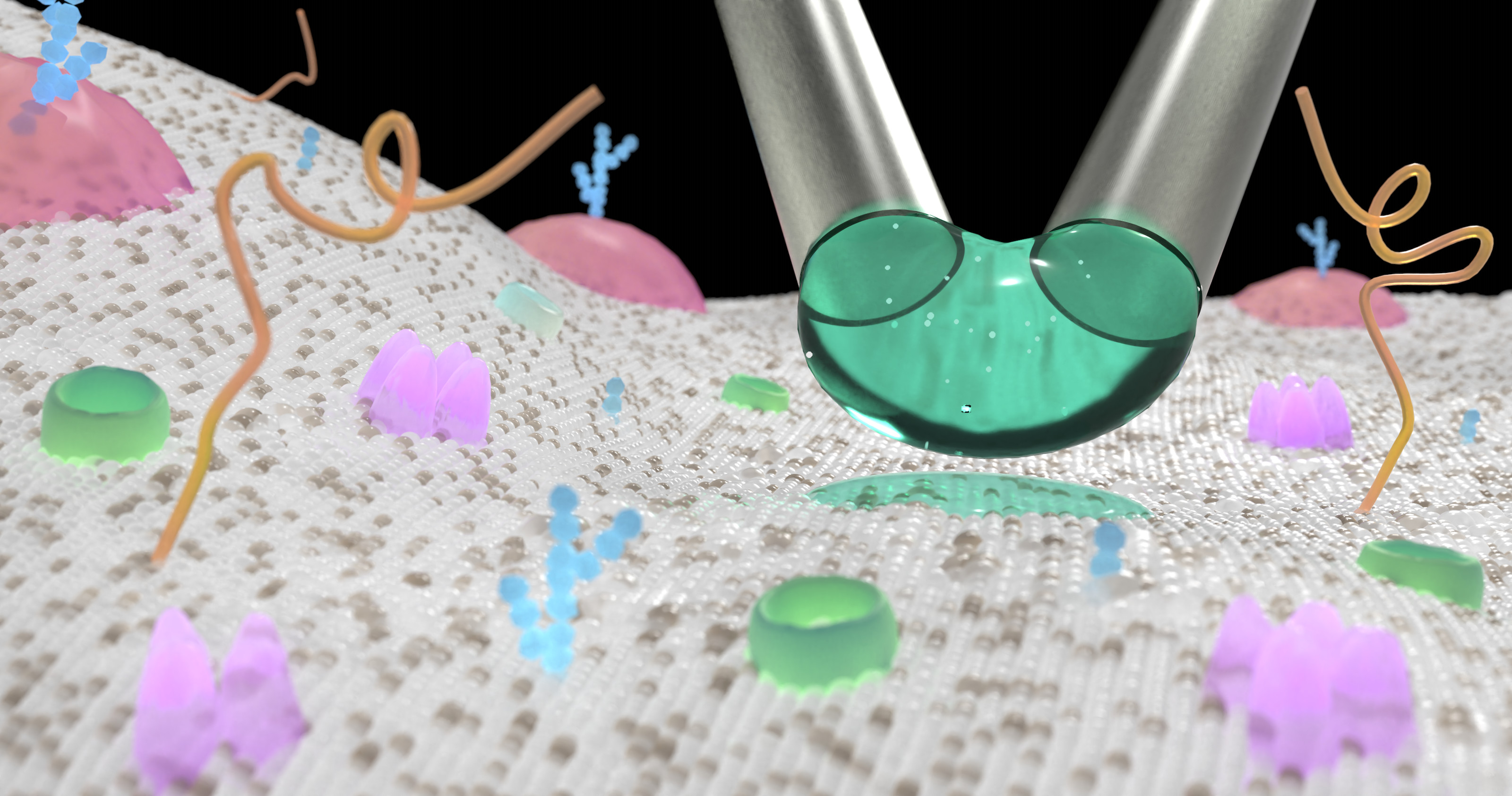
A droplet can be positioned with micropipettes and gently brushed against a cell. This releases a tiny “μkiss” (© MPL, Dr. Richard W. Taylor).
Traditional approaches in biology often consider characteristics across entire cell populations, missing the nuanced variations in properties from one cell to another. To investigate biology more precisely at the individual cell level, the development of new tools and methods is imperative. “A crucial gap remains in our ability to administer chemicals, labels, and pharmaceuticals to individual cells with precision and control, over short durations and miniscule microscopic length scales”, says Professor Vahid Sandoghdar, Director of the Max Planck Institute for the Science of Light and Max-Planck-Zentrum für Physik und Medizin. Prof. Sandoghdar and his team have been actively addressing this challenge.
"Like a paintbrush", easy and cost-effective to use
The researchers devised a simple yet elegant solution to this problem: using two closely placed micropipettes with an opening as small as just one micrometer, the scientists could create a stable micro-sized droplet of material at the micropipette ends by using one micropipette to dispense the material, while the other suctions it in at a slightly higher rate. “It is then just like a paintbrush”, says Richard W. Taylor, Post-doctoral researcher and member of the team, adding “You can easily maneuver the micropipettes around, and gently brush this confined droplet against your chosen cell – delivering a tiny μkiss of material”.
This simple implementation, using readily available components allows their technique to be easily implemented at low cost on any microscope within biologically-orientated laboratories. "The cost-efficient and pragmatic approach of our solution is important for its use in practice”, says Prof. Sandoghdar, adding "The lack of similar solutions has so far delayed progress towards new therapeutic approaches at the single-cell level“.
Full control over location, time and scale
The new method places the experimenter in full control. “With μkiss, we achieve a completely new dimension in the precise application of substances to cells”, explains Cornelia Holler, a doctoral student in Biology and member of the research group. Materials can now be precisely delivered to any chosen cell at the sub-cellular level, with complete control over the time and position the material is in contact with the cell. “We can now watch entire biological processes, such as the uptake of iron by the cell, without missing a step – this allows us to finally piece together the puzzle of the complex characteristics of each individual cell," says Holler.
Recently, the team achieved the precise placement of a single virus-like particle onto a live cell. This experimental ability creates an opportunity to scrutinize the intricacies of disease propagation, providing full control over the location, timing, and extent of cell infection. “The ability to μkiss opens new pathways for quantitative studies in cell biology and Medicine”, says Professor Sandoghdar.
Original publication in Nature Methods
Holler, C., Taylor, R.W., Schambony, A. et al. A paintbrush for delivery of nanoparticles and molecules to live cells with precise spatiotemporal control. Nat Methods (2024).
DOI: https://doi.org/10.1038/s41592-024-02177-x
Scientific contact:
Sandoghdar Division
Max-Planck-Institut für Physik und Medizin, Erlangen
mpzpm.mpg.de / sandoghdar-office@mpl.mpg.de
Contact
Edda Fischer
Head of Communication and Marketing
Telefon: 09131 7133 805
MPLpresse@mpl.mpg.de